Every year in honor of National Nanotechnology Day on October 9th, the National Nanotechnology Coordinated Infrastructure (NNCI) hosts a Plenty of Beauty at the Bottom image contest to celebrate the beauty of the micro and nanoscale.
The Northwest Nanotechnology Infrastructure, of which the Institute for Nano-engineered Systems leads, is one of NNCI’s 16 member sites. Researchers are invited to submit images of their work taken at NNCI facilities (which at the UW includes the Washington Nanofabrication Facility and the Molecular Analysis Facility) for consideration for the categories of Most Stunning, Most Whimsical and Most Unique Capability.
The winners will be entered into a network-wide competition. Public voting begins October 8.
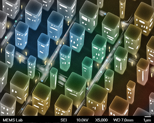
Most Whimsical – Winner
Micro-City
Zheyi Han, Electrical & Computer Engineering Graduate Student in the Bohringer Lab
Taken on a JEOL-JSM7400F Scanning Electron Microscope at the Washington Nanofabrication Facility
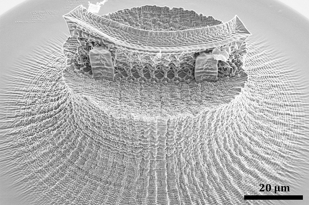
Most Stunning – Winner
Nano-wrinkled Head
Zainab Patel, Materials Science & Engineering Graduate Student in the Meza Lab
Taken on an Apreo SEM in the Molecular Analysis Facility
2021 Featured Submissions

An image of an electrochemical electrode array fabricated at the WNF facility was superimposed with a picture of a boat. The pattern is formed of 50 micron-wide lines.
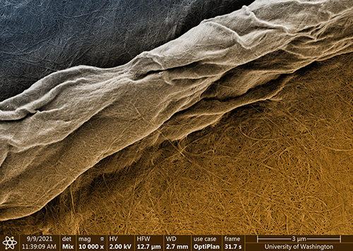
This image depicts a cellulose macrofiber isolated from plants and its incredible hierarchical structure, leading to much smaller, individual microfibers that spread over the image’s background. The bigger macrofiber is a few microns wide, while the smaller ones are just tens of nanometers wide. We were very excited to simultaneously capture fibers at two very different scales (micro and nano) in a single frame.

An image of electrochemical electrodes fabricated at WNF by postdoc Tran Nguyen (Folch lab, UW BioE) has been distorted for perspective and overlaid with that of a lagoon and a boat.
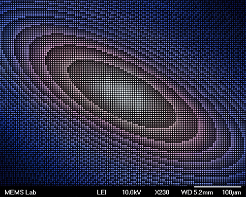
False-colored scanning electron microscopic image of silicon pillars arranged as part of a micro-lens.
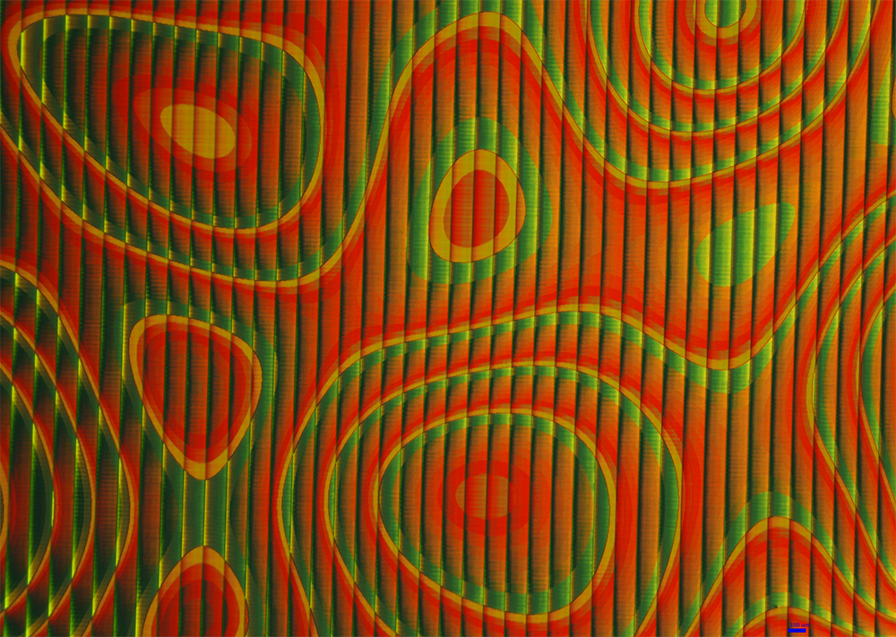
Several tens of thousands of micro pillars (in the micrometer scale) with slightly varying dimensions are distributed very close to each other in a specific layout producing this weird shape. The micro structures are made of a photosensitive resin, and diffract different colors according to their sizes. The vertical stripes are due to a non uniform exposure to light. The specific shapes are meant to redirect light rays in different directions to recreate an optical neuron-like network.
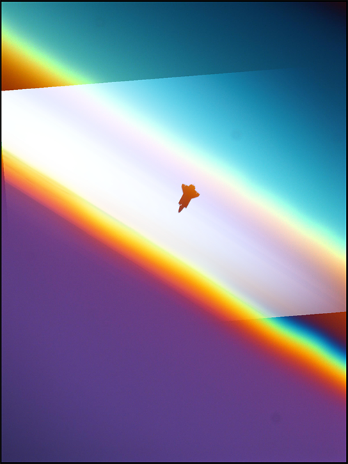
Combination of image of a thin oxide film and image of a space shuttle in space.
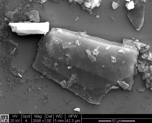
Black phosphorus flake with multiple layers as seen by scanning electron microscopy. As a two dimensional material, black phosphorus can be exfoliated down into single and few layer sheets consisting of only phosphorus atoms similar in many ways to graphene (a two dimensional material consisting solely of carbon atoms).